


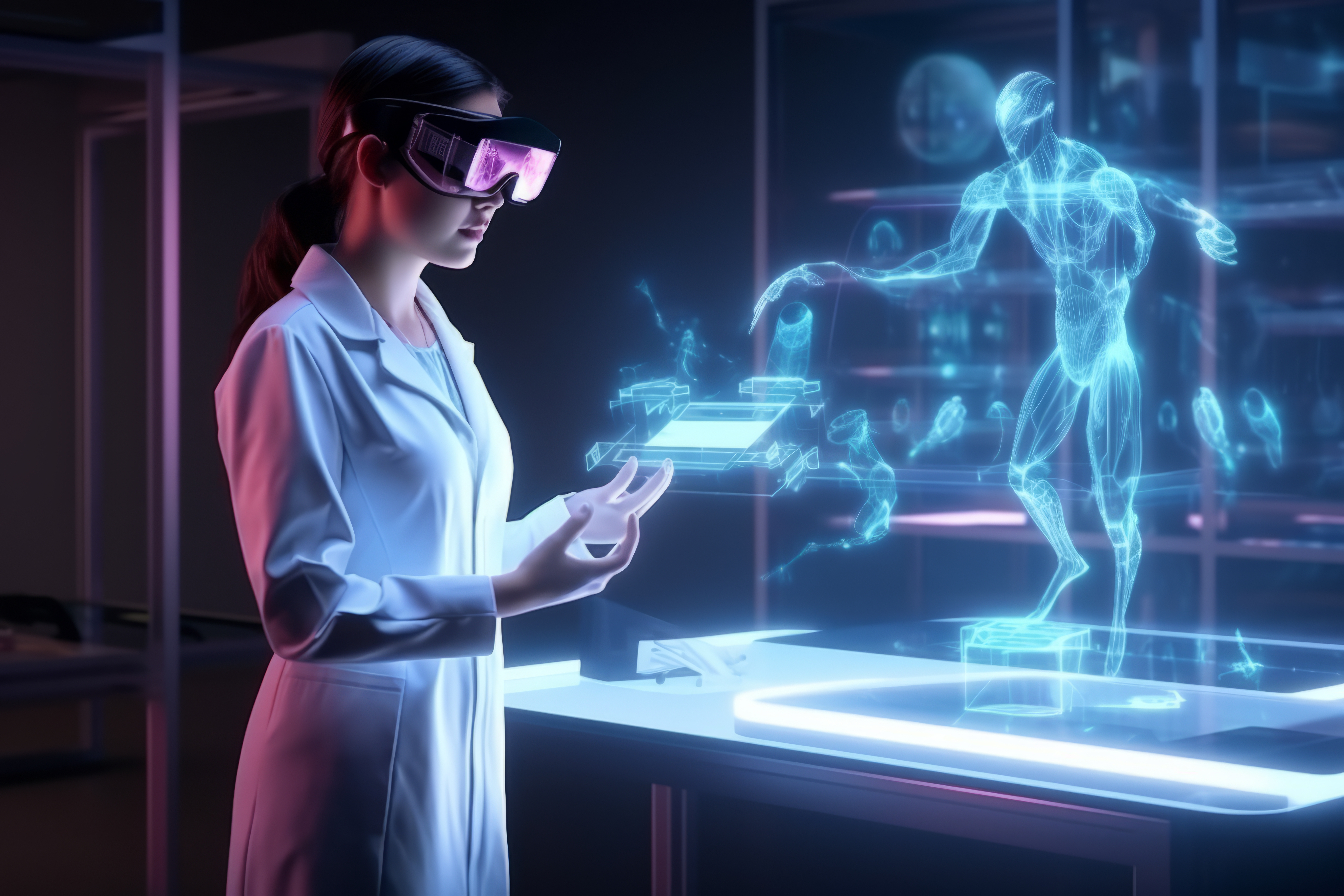
Deo Anatomy - VR Anatomy
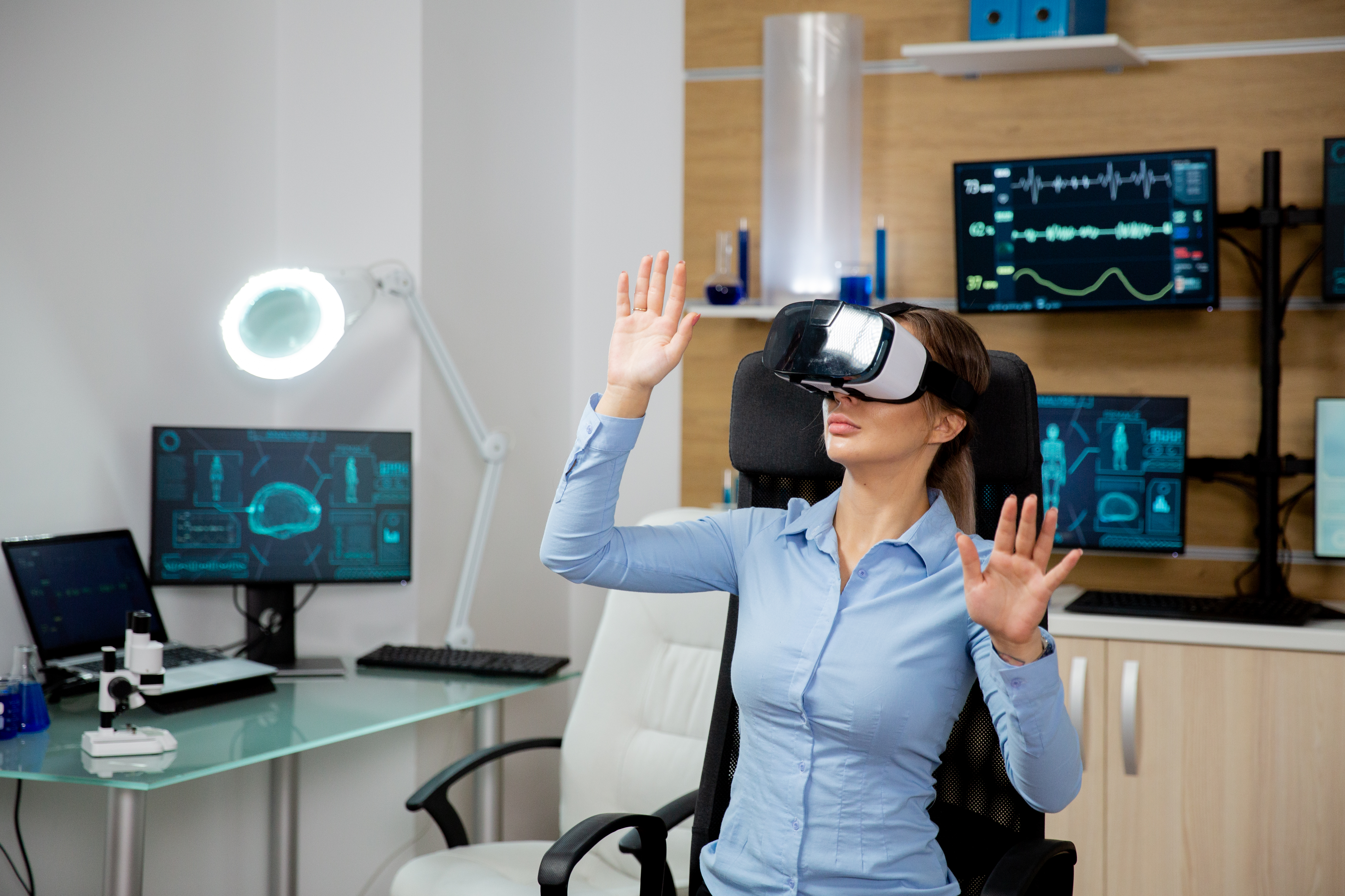
Deo Anatomy a VR Anatomy Software is an advanced educational tool designed to teach human anatomy in an immersive, interactive Virtual Reality (VR) environment. Unlike traditional methods of studying anatomy using textbooks or 2D models, VR Anatomy Software allows students, medical professionals, and even surgeons to explore detailed 3D representations of the human body. This technology is revolutionizing how anatomy is taught and learned by offering a more hands-on, engaging, and realistic experience.
Here’s a detailed explanation of what VR Anatomy Software entails:
1. Immersive 3D Exploration of Human Anatomy
The primary feature of VR Anatomy Software is its ability to provide a fully immersive, 3D environment where users can explore the human body in real-time.
-
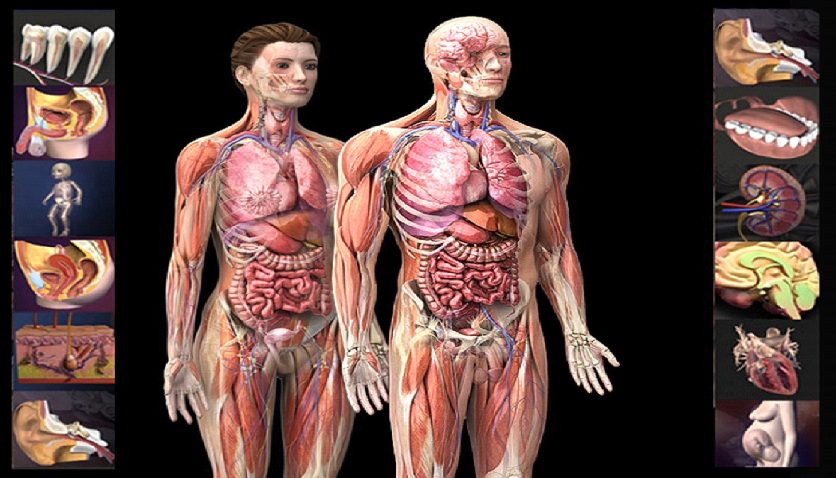
Detailed Visualization: The software presents anatomically accurate 3D models of every organ, muscle, bone, and system within the human body. Users can zoom in, rotate, and examine each component from any angle, offering a far more detailed view than what’s possible with 2D diagrams or even physical cadavers.
-
Layered View: Users can peel back layers of the body to view different systems, such as the skeletal system, muscular system, circulatory system, nervous system, and more. This layered approach allows for a comprehensive understanding of how each part of the body interacts with others.
2. Interactive Learning
-
Manipulation of Structures: Users can manipulate and isolate specific body parts, such as bones, muscles, organs, or tissues. For instance, they can remove the heart from the body, enlarge it, and examine its internal chambers.
-
Exploration Tools: The software typically includes tools for slicing through tissue, enabling users to see cross-sections of organs or systems. For example, they can perform a virtual dissection to study the inner structures of the brain or heart.
One of the biggest advantages of VR Anatomy Software is the ability to interact with the anatomy in ways that are not possible with traditional tools.
3. Realistic Medical Training
-
Simulated Procedures: Students can simulate medical procedures or dissections within the VR environment, allowing them to practice without the need for cadavers. This simulation helps build essential skills in anatomy, surgery, and diagnostics.
-
Dynamic Learning: VR anatomy offers real-time feedback, with quizzes, annotations, and guided learning pathways integrated into the software. This enhances knowledge retention and makes the learning process more dynamic.
VR Anatomy Software is widely used for medical education and training, allowing students and professionals to practice and refine their understanding of human anatomy.
4. Enhanced Understanding of Complex Anatomy
-
Exploring Complex Areas: For example, the software allows users to explore the nervous system in 3D, following nerve pathways from the brain down to the extremities. This is particularly useful for understanding how different systems are interconnected.
-
Pathology and Conditions: Many VR Anatomy platforms allow users to visualize abnormal anatomy, such as tumors, injuries, or diseased organs. This feature is vital for medical students and professionals studying pathology, as it helps them understand the physical manifestations of disease.
VR Anatomy Software excels at teaching complex anatomical structures and systems, such as the brain, cardiovascular system, and muscles.
5. Collaborative Learning
-
Shared Learning Spaces: Medical students or professionals in different locations can enter the same VR environment and explore anatomy together. This feature is particularly useful for group study, medical conferences, or consultations between medical professionals.
-
Instructor-Guided Sessions: Educators can lead guided sessions in VR, pointing out important structures or explaining complex systems to their students in an interactive, 3D space.
VR Anatomy Software allows multiple users to interact with the same virtual anatomy in real-time, making it a collaborative tool for learning.
6. Practical Applications in Surgery and Medicine
-
Preoperative Planning: Surgeons can use the software to explore a patient’s anatomy in VR before an operation. This allows them to visualize surgical pathways, plan procedures, and identify potential complications.
-
Patient Education: Doctors can use VR anatomy to explain conditions and treatments to patients, helping them better understand their medical situation. For example, a doctor could show a patient their heart in 3D, explaining how a procedure like a bypass surgery will be performed.
For practicing surgeons and medical professionals, VR Anatomy Software can serve as a crucial tool for surgical planning and patient education.
7. Scalable for Different Fields of Medicine
-
General Anatomy: Suitable for medical students, nurses, and healthcare professionals who need a foundational understanding of the entire human body.
-
Specialized Fields: The software can focus on specific fields, such as cardiology (heart anatomy), neurology (brain anatomy), or orthopedics (skeletal system), making it versatile for advanced medical training.
-
Surgical Simulation: Surgeons can use the software for procedure-based training, such as practicing surgical approaches or studying the anatomy of a particular patient before surgery.
VR Anatomy Software can be tailored to various medical specialties, including:
Deo Simulation - VR Simulation for Medical Trainees
Our VR simulations provide an immersive environment where medical students and trainee doctors can practice procedures and study anatomy in a highly realistic, risk-free virtual world. The goal is to bridge the gap between theoretical knowledge and real-world practice, allowing trainees to build confidence before stepping into real clinical environments.
Here’s a detailed explanation of what VR Anatomy Software entails:
a. VR Anatomy Software
This cutting-edge software offers a highly interactive and detailed 3D representation of human anatomy. Medical students can:
-
Explore every layer of the human body, from bones and muscles to organs and tissues, in lifelike 3D detail.
-
Dissect and manipulate anatomical structures for a hands-on learning experience, without the need for cadavers.
-
Visualize complex systems such as the cardiovascular, nervous, and skeletal systems in an interactive, dynamic way.
-
Use VR-enabled learning modules to understand how various body systems interact, enhancing the comprehension of physiological processes.

b. VR Surgery Simulation for Bachelor of Medicine, Bachelor of Surgery (MBBS)
-
Realistic surgical scenarios where students can perform procedures like appendectomies, heart surgeries, and more.
-
Interactive tools that replicate real-world surgical instruments, offering a hands-on experience.
-
Detailed feedback systems that track precision, timing, and technique, allowing trainees to refine their skills.
-
Collaboration tools enabling students to work in teams, simulating real-world surgical teams and operating room dynamics.
This simulation platform allows MBBS students to practice complex surgical procedures in a controlled, virtual environment. Key features include:
c. VR Surgery Simulation for Bachelor of Dental Surgery (BDS)
-
Virtual patients with realistic dental anatomy, where students can perform tasks such as root canals, extractions, and dental implant procedures.
-
Simulation of tools like dental drills, forceps, and scalers to provide a tactile experience through VR controllers.
-
Feedback mechanisms that assess the student’s accuracy and technique, allowing continuous improvement in a safe environment.
-
Enhanced learning modules focusing on various dental conditions and treatment plans, preparing students for real-world patient interactions.
Tailored specifically for dental students, this simulation offers real-life scenarios for practicing dental surgery and procedures. Key features:
3. Deo Vision - Dicom Viewer
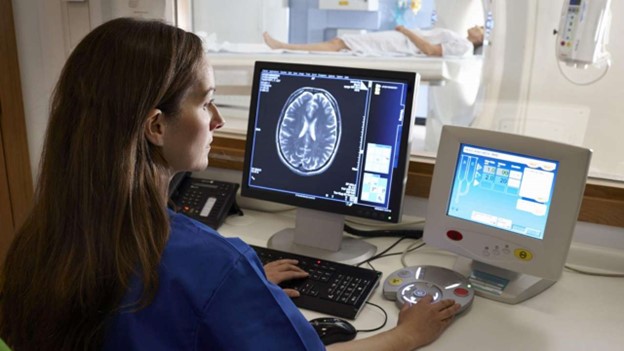
Deo Vision VR Viewer is a cutting-edge medical application that allows healthcare professionals to view, interact with, and analyze DICOM (Digital Imaging and Communications in Medicine) files in a fully immersive Virtual Reality environment. DICOM files are the standard format for storing medical imaging data, such as MRI, CT scans, ultrasound, and X-rays, and are crucial for diagnosis, surgical planning, and treatment.
Here’s a detailed explanation of what VR Anatomy Software entails:
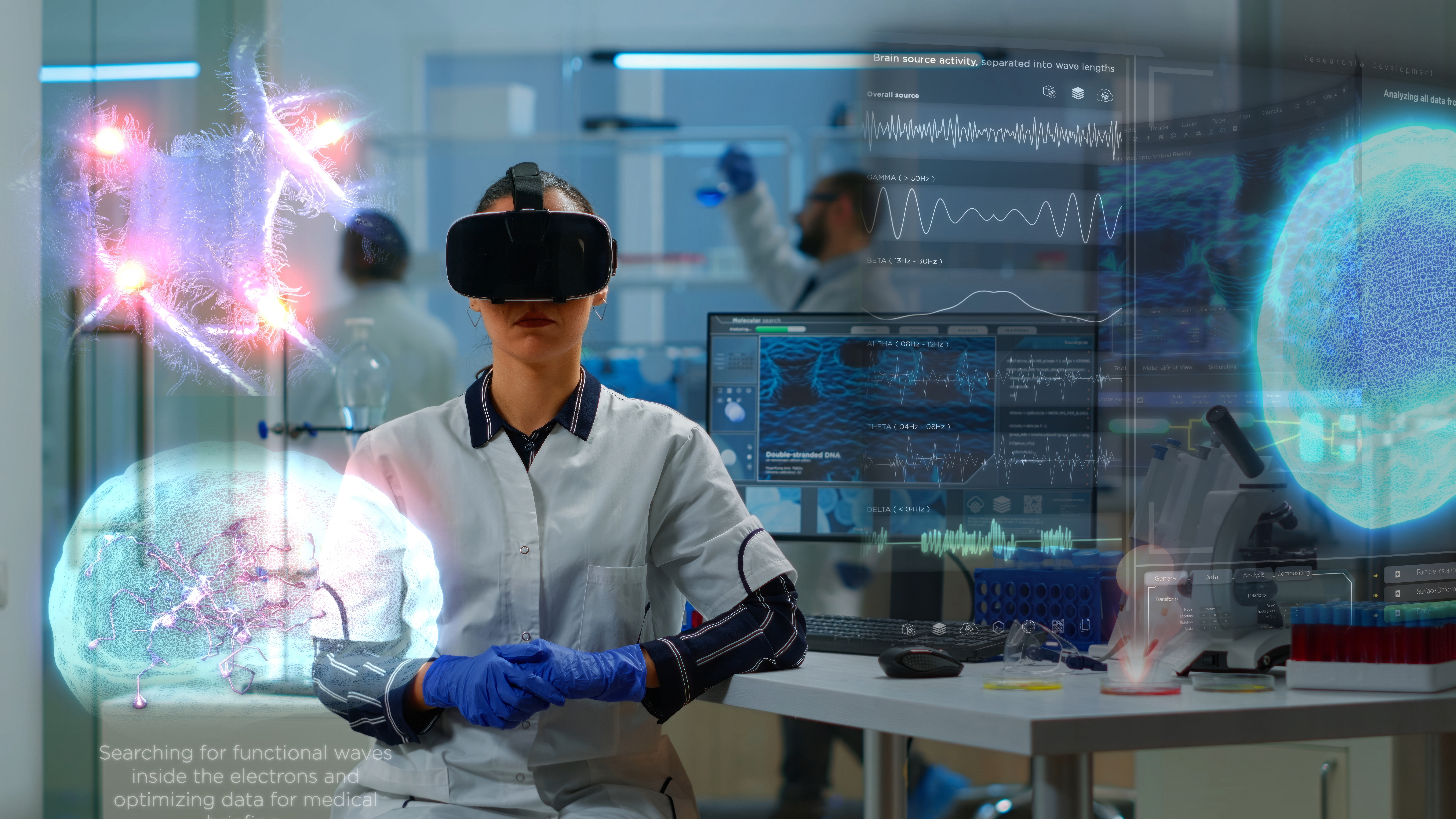
1. Immersive 3D Visualization
The primary feature of a DEO Vision is its ability to transform traditional 2D medical images into fully immersive 3D models. Instead of viewing flat images on a screen, doctors can step into a virtual environment where they can interact with the medical images as though they were physical objects. This allows for a much more detailed examination of complex anatomical structures, tumors, blood vessels, and organs.
-
3D Reconstruction: The viewer can reconstruct 2D DICOM data into 3D models, enabling medical professionals to rotate, zoom in, slice, and explore anatomical structures in real time. This capability is particularly useful for understanding the spatial relationships between different parts of the body.
2. Enhanced Diagnostic Capabilities
-
Detailed Exploration: Physicians can explore multiple layers of anatomy and adjust settings such as opacity to focus on specific tissues or structures (e.g., bones, muscles, or organs).
-
Better Diagnostic Insight: The immersive environment allows doctors to view images from any angle, improving diagnostic accuracy and potentially identifying conditions that might be missed in 2D viewing.
By utilizing VR, medical professionals can gain a deeper understanding of the patient’s anatomy, which enhances diagnostic accuracy and treatment planning.
3. Surgical Planning and Simulation
-
Preoperative Planning: Surgeons can use VR to walk through the patient's anatomy before performing surgery, giving them a clearer understanding of what they will encounter in the operating room. For example, they can practice navigating around delicate structures such as nerves and blood vessels.
-
Simulating Procedures: By overlaying DICOM data in VR, surgeons can practice certain surgical approaches or techniques, visualizing potential complications and adjusting their plans accordingly.
A Deo Vision Viewer is highly valuable for surgeons who need to prepare for complex procedures.
4. Collaboration and Remote Viewing
-
Remote Collaboration: Physicians from different locations can share and discuss DICOM data in real time, viewing and manipulating the same 3D images together. This is particularly beneficial in telemedicine, multidisciplinary consultations, or training environments.
VR allows for a collaborative approach to medical imaging. Multiple doctors, surgeons, or medical students can enter the same virtual environment, enabling collaborative diagnosis, treatment planning, or even teaching.
5. Teaching and Education
-
Interactive Learning: Students can explore complex medical images in 3D, improving their understanding of human anatomy and various medical conditions. The immersive aspect helps bridge the gap between textbook learning and real-world clinical experience.
-
Practice Interpretation: Trainees can practice interpreting DICOM images in VR, enhancing their radiological skills and preparing them for real-life clinical scenarios.
For medical students and trainees, the Deo Vision Viewer offers an interactive and immersive way to learn about anatomy, radiology, and pathology.
6. Improved Patient Communication
-
Patient Education: Physicians can use VR to show patients a 3D visualization of their condition, helping them better understand their diagnosis and the proposed treatment plan. This improves patient trust and engagement in their healthcare.
Deo Vision also benefit patient care by making it easier to explain complex medical conditions and treatments to patients.
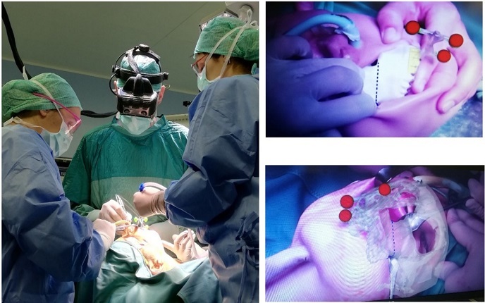
4. Deo Surgery - Augmented Reality for Real Time Surgery
Our AR solutions provide a revolutionary way for surgeons to enhance their precision during complex surgeries by overlaying critical data in real time. Our AR technology integrates MRI, CT scans, and other imaging data, converting them into 3D visualizations that can be superimposed onto the patient's body.
Key Features:
-
Real-Time 3D Visualizations: Surgeons can view 3D models of the patient’s anatomy in real time, directly overlaid onto the patient’s body. This ensures that the surgeon has a clear understanding of where vital organs, blood vessels, and potential risks are located.
-
Improved Surgical Accuracy: By superimposing imaging data onto the patient’s body during surgery, our AR solutions help reduce errors, improve precision, and ultimately lead to better patient outcomes.
-
Seamless Integration with Medical Imaging: Our AR devices are fully compatible with modern medical imaging technologies, including MRI and CT scans. This allows surgeons to access real-time data during surgery without disrupting the workflow.
-
User-Friendly Interface: Surgeons can interact with the AR device through simple hand gestures or voice commands, keeping the interface intuitive and easy to use during critical moments.
-
Applications in Various Surgical Fields: Whether it's neurosurgery, orthopedics, or cardiovascular procedures, our AR technology can be customized to meet the specific needs of various surgical specialties.
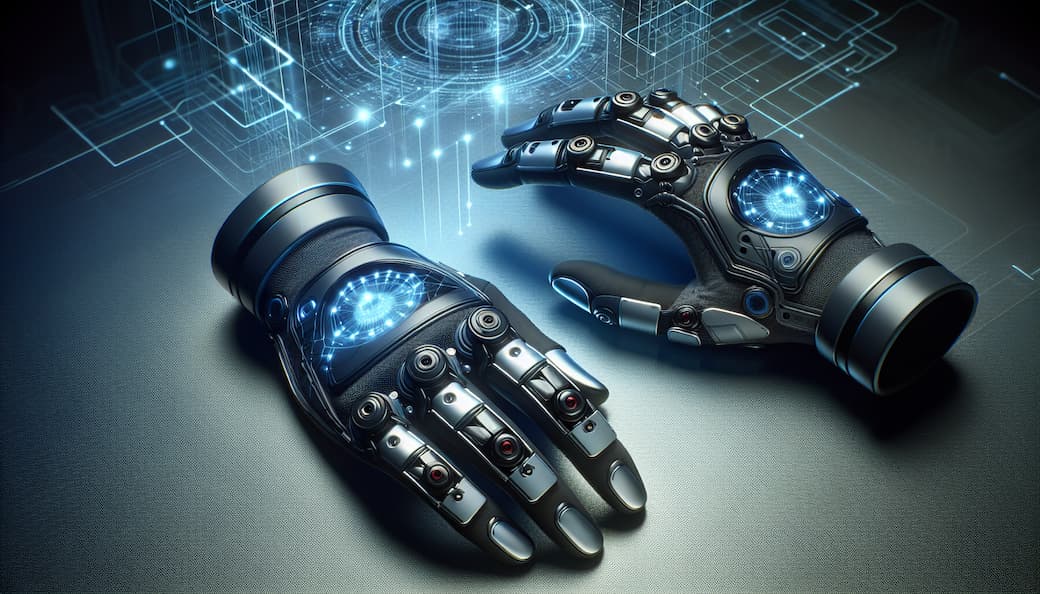
4. Deo Gloves – Haptic Gloves for Medical Simulation
One of the biggest challenges in virtual reality training is the lack of tactile feedback. Our haptic gloves are designed to solve this by providing a realistic, tactile experience, allowing medical professionals to feel the textures, pressure, and resistance of tissues, organs, and instruments in a virtual environment.
Key Features:
-
Realistic Tactile Feedback: The gloves simulate the sensation of touch, allowing users to feel virtual objects as though they were physically present. Surgeons can feel the texture, density, and pressure required for each virtual surgical task.
-
Enhanced Training Experience: Trainees can practice complex procedures like suturing, incisions, and organ manipulation with a highly realistic sense of touch. This helps them refine their motor skills and get a feel for the procedures before performing them in real surgeries.
-
Pressure Sensitivity: The gloves detect and simulate varying degrees of pressure, helping trainees understand the force needed when interacting with tissues or surgical instruments.
-
Applications Beyond Surgery: While the primary focus is on surgery, these gloves can also be used for other medical training scenarios, such as palpation, diagnostic assessments, and even robotic surgery simulations.
-
Ergonomically Designed: The gloves are lightweight and comfortable, allowing extended use without discomfort or fatigue, ensuring a seamless training experience.